Pathophysiology of Vaccine-preventable diseases: Part 3

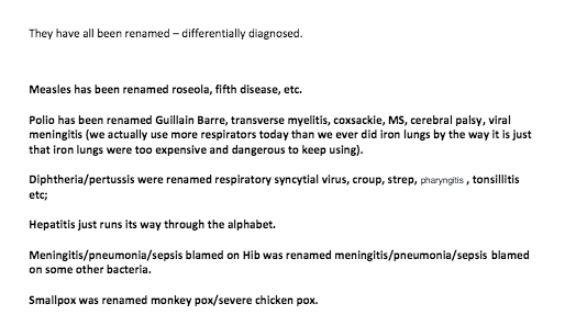
Part 3 is specifically made for Tristan Wells, angry Australian protestor against vaccination and Germ Theory who may or may not have the IQ of a humpback whale.

Normally I’d feel bad for singling out someone on the internet,  but I can’t quite express how communicating with him feels somewhat like talking to a very angry brick wall. Mistakes were made. Here is an excerpt from his blog on how to debate a pro-vaxxer:

Screen Shot 2018-04-19 at 8.56.14 PM

I know what you’re thinking. “Oh come on, he’s trolling.”

He isn’t. He’s serious. He doesn’t believe in Germ Theory in 2018 and thinks all diseases (I don’t know what he thinks diseases come from either) are renamed. Not just confused with other conditions back in ancient times (which is plausible), or differential diagnoses (which is taken into consideration and part of every patient’s paperwork), but secretly renamed other conditions to convince the world that vaccines work. Welcome to anti-vaxx nutville 2018.

Here is why we’re not “renaming” organisms:

Pathogens have been structurally observed and drawn out. Even if you can’t directly observe it, the body will still create monoclonal antibodies against that organism specifically, not against an organism that has a similar clinical presentation. Vaccine preventable diseases, tetanus being the exception, are not diagnosed by presentation alone but serology, cell culture, or anther specific diagnostic test. Tetanus is the exception because only a few organisms are needed to initiate an infection and they’re obligate anaerobes (exposure to O2 will kill them), making it extremely difficult to isolate. And also because it’s really fucking obvious if patient has lockjaw.

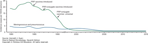
If meningitis, pneumonia, and sepsis were being renamed, it’s amazing how S. pneumoniae is blatantly mentioned in all of my board prep material as the most common cause of community acquired pneumonia and H. influenzae the most common cause of acute epiglottitis. Whenever he’s trying to argue (I’ve had the pleasure of accidentally talking to him once on twitter), he always says what doctors do and don’t do for differential diagnoses, and is wrong every time. He will flip out when you tell him that’s not how it works. He’s convinced that doctors will ignore signs and symptoms of a vaccine preventable disease in vaccinated individuals, which isn’t even close to what actually happens. Signs and symptoms means just that: signs and symptoms. On top of it, we’re obviously not going to have someone’s entire vaccine history in a hospital, but we’ll add it to a SOAP note if they’re not actively dying and we can ask them in person. Pneumococcal pneumonia and Hib are still up there as expected diagnoses of CAP and meningitis. Sepsis can be caused by many different organisms such as E. coli, and they’re going to test for it. If someone who is young and vaccinated comes in with signs of pneumonia and doesn’t have to be admitted, do you know what we treat them for? You guessed it. Without testing, we automatically assume it’s S. pneumoniae and treat them with macrolides. We are more likely to assume S. pneumoniae than whatever other infection Tristan is thinking we’re trying to secretly sell (or whatever he thinks we’re doing), even though he also somehow simultaneously doesn’t believe in Germ Theory.

Fifth Disease vs. Rubeola (measles) vs. Roseola (and I’ll even throw in HFM for free!)

Parvovirus B19 (fifth disease)

Virus

Genome

Disease

Transmission/Dx

Tx/Vaccine

Comment

Parvovirus

B19 (fifth disease) (Erythema infectiosum)

ssDNA

naked

linear

1/5 childhood rashes; must infect replicating cells (erythroid)

trasmission: oral, respiratory droplets;

Dx: IgM IgG serology; PCR for DNA

no antivirals available; vaccine available for animals only

most adults sero+; resistant naked capsid; shed virus before symptoms; crosses placenta

Measles aka Rubeola

Virus

Genome

Disease

Transmission/Dx

Tx/Vaccine

Comment

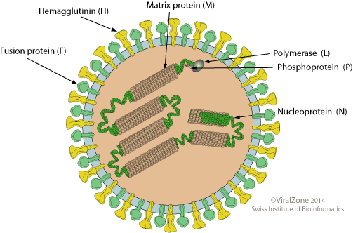
Paramyxoviridae

Morbillivirus

Measles AKA rubeola

ssRNA (-) enveloped

measles -> pneumonia, encephalitis, subacute SSPE, CCC&P;

aerosols/

Dx: giant cells from fusion

Vaccine: MMWR (live)

Tx: no antiviral

humans = host;

Unique features: maculopapular rash & Kopliks spots; replicate in RT but spreads thru blood -> disease; can spread cell-cell, avoids Abs

HHV-6 aka Roseola

Virus/Family

Genome

Disease

Transmission/Dx

Tx/Vaccine

Comment

Herpesveridae

Betaherpesviridae

Human Herpesvirus 6

AKA Roseola

dsDNA;

enveloped linear

Appears as prodrome sequence; acute high fever (104 F) followed by classic rash with no other symptoms

Aerosols, saliva, direct contact

self-limiting, CMI

HHV6 also called Roseola or exanthema subitum; rash from activation of Tcells

Coxsackievirus (not synonymous with HFM, which is a clinical presentation)

Virus

Genome

Disease

Transmission/Dx

Tx/Vaccine

Comment

Picornaviridae

Coxsackievirus A

+ssRNA

naked

herpangina; hand foot & mouth; encephalitis, meningitis; carditis, common cold, & more

fecal-oral

Dx: fecal culture; CSF has low neut

pleconaril if early

differ strains -> differ disease; infants highest risk;

Picornaviridae

Coxsackievirus B

+ssRNA

naked

pleurodynia, encephalitis, meningitis; carditis, common cold, & more

fecal-oral

Dx: fecal culture; CSF has low neut

pleconaril if early

differ strains -> differ disease; infants highest risk;

Key differences:

  • He managed to pick organisms that had completely different nucleic acids from one another. This isn’t like he picked related herpesviruses and asked why we don’t usually get confused – he picked viruses that aren’t remotely similar. All he needs now is a retrovirus and he covered about all of the bases
  • HHV-6 will have no other symptoms
  • Why would we even “rename” fifth’s with sixth’s disease when neither one of them has a vaccine?


Rashes:

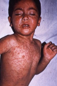
Childhood rashes are distinct from one another.
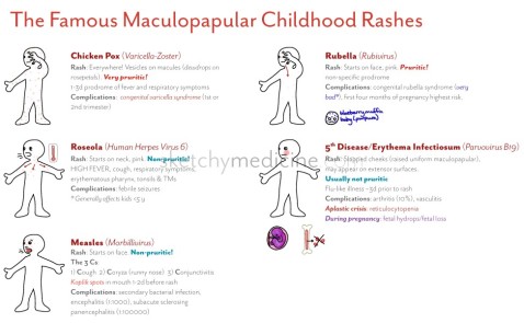
rashes.jpg

VARICELLA:

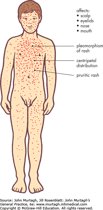
  • The pruritic lesions appear in “crops” for about 3-4 days
    • The lesions are characteristically described as “dew drop on a rose petal”
    • Each lesion is considered contagious until a scab forms
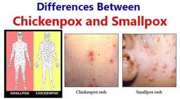
    • The “chickenpox” are at different stages of healing 
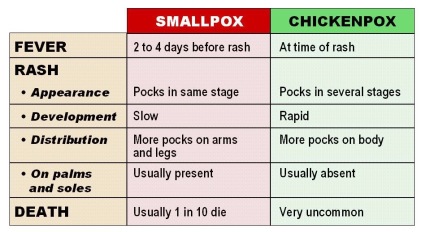
      • This sets it apart from smallpox – smallpox will never be at different stages of healing! 
  • Spread: From face/trunk to extremities

    Duration: ~1 week

ROSEOLA:

  • Diffuse, splotchy, maculopapular rash that is NOT pruitic
  • This automatically separates it from chickenpox 
    • Rash duration is approximately 24 hours
      • Shorter rash duration than the others
    • leave no scars — unlike chickenpox

    Spread: Very quick – Trunk to extremities and to

    entire body


Strep– This is caused by Streptococcus pyogenes. Strep throat IS tonsillitis caused by S. pyogenes. Tonsillitis can be caused by multiple different organisms. Yes, sometimes including vaccine-preventable diseases. This is why we test patients and include multiple differentials. And yes, tristan, we can easily tell a difference.

The general appearance alone can let someone know they’re dealing with strep or diphtheria. However, patients are still going to be swabbed and tested.


Diphtheria- pseudomembrane located on throat
Strep throat progression – no pseudomembrane, located on tonsils


The same goes for pharyngitis and tonsillitis.

Signs and symptoms with variable causes such as pharyngitis and tonsillitis do not determine pathogen upon diagnosis, but rather a disease presentation. To put this in the explain-it-like-im-five version, If you see “-itis,” that means inflammation. So meningitis means an inflammation of the meninges. Pharyngitis is an inflammation of the pharynx, and arthritis is an inflammation of the joints. Some definitions are even more vague, like myalgia. That’s not a disease, that’s a presentation with a slew of different etiologies. That simply means muscle pain. Confusing a pathogen with a presentation is like confusing Van Gogh, the person, with a presentation like impressionism. It doesn’t make an ounce of sense. Streptococcus may be the main etiological agent of pharyngitis, but we still test it if a patient comes in.


Whooping cough vs literally every other cough in existence:

How do we know it’s whooping cough and not a different illness?

For starters: “COUGH COUGH COUGH COUGH COUGH COUGH cough cough cough cough cough cough (x 30) WHOOP” for 100 days kinda gives it away.

The clinical presentation and cough are very specific. The first step in the official diagnosis of pertussis is to have suspicion and proper testing:

  • patient with a paroxysmal cough for 14 days or longer and one or more of the following criteria:
    • paroxysmal cough, whoop, or posttussive vomiting.
    • PCR assay or a confirmed epidemiologic link.

Yes, you are going to be tested for it.  Yes, Tristan, even vaccinated kids

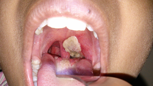
Diphtheria can have a variable presentation, so how do we know it’s not a different infection? How do we know it’s not streptococcal or viral pharyngitis or tonsillitis, or Vincent’s angina?

AGAIN: BY TESTING FOR IT!  

This information is straight out of my evil Big Pharma class notes

Diphtheria should be considered in any patient with the following symptoms:

  • tonsillitis and/or pharyngitis with pseudomembrane
  • hoarseness and stridor
  • cervical adenopathy or cervical swelling (bull neck)
  • unilateral bloody nasal discharge or paralysis of the palate

Ruled out by culture and isolation

Different structures:

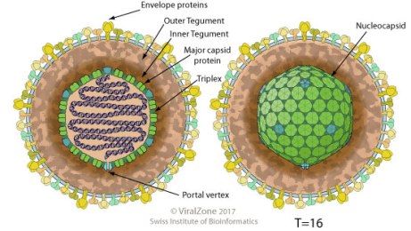
General structure of Herpesviruses:

Herpesviridae_virion.jpg

These have similar structure and nucleic acid. This is why they’re in the same family. This doesn’t mean they’re the same disease, this means you wouldn’t diagnose it with an electron microscope (which you wouldn’t do anyways). They have completely different presentations and pathogenesis

vs another DNA virus like:

SMALLPOX

Poxviridae_virion.jpg

They are a completely different shape, and smallpox is also very large and can technically be seen with a light microscope

Monkeypox vs smallpox-

Smallpox

Virus/Family

Genome

Disease

Transmission/Dx

Tx/Vaccine

Comment

Poxviridae

Smallpox aka Variola

linear dsDNA

URT & 2nd viremia->rash; prodromal fever then centrifugal, regular rash

fomite or lesion/ Guarnieri body

Vaccine available; VIG 1st; Cidofovir 2nd

very contagious, can go through skin; humans only host

Monkeypox

Monkeypox

poxviridae

linear dsDNA

similar to smallpox, reappearing due to vaccinia

african squirrel-> rodents

US outbreak 2003 from imported rodents & housepets

Monkeypox is a zoonotic infection that can appear similar at first to smallpox, which would have scared the living hell out of me if I saw that for the first time. But they’re not the same. Sorry, Tristan. Again, to repeat from Part 2: You diagnose smallpox via both the clinical presentation and diagnostic tests testing specifically for the DNA. You would never ever ever ever ever (continued 100x) have a clinical presentation of smallpox without knowing for certain that the person does or doesn’t have smallpox by running specific diagnostic tests. I have to say this because he’s convinced we don’t add vaccine-preventable diseases as part of our differential. And not only would we, but we would be REALLY FUCKING SURE we weren’t dealing with smallpox. Even if you took the most selfish doctor on earth who didn’t care about their patients, they don’t exactly want smallpox either and would report it to save their own ass and their own literal skin. There is no reason to hide a smallpox diagnosis that wouldn’t result in an immediate firing, a lockdown of a hospital, and a country freaking out. Nothing good would come out of hiding a smallpox diagnosis.


Monkeypox

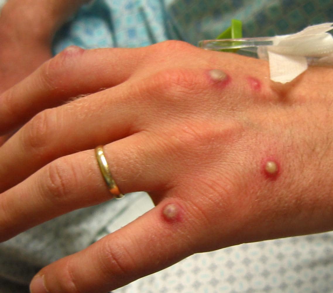
First of all, monkeypox is still extremely rare. Tristan never really explained how so many contracted and died of smallpox every year, and a few people here and there have been affected by monkeypox. Weird… where did they all go? Even if his opinion was true (which we all know it’s not), he still doesn’t explain why there was a sudden drop in smallpox to monkeypox. Wow…. weird.

Monkeypox vs smallpox won’t be diagnosed from observation and biopsy. That’s because both involve a similar morbiliform exanthem. Monkeypox in a clinical presentation that’s milder than smallpox and involves a veroliform rash with progression from papules to vesicles, umbilicated pustules, and crusting. We test it with PCR, ELISA, and/or Western blotTristan will say “So? How do you know 100% of smallpox cases were smallpox in the 1600s?” He still doesn’t realize that not only is this a stupid argument (he’s asked this), but it still doesn’t explain how monkeypox was so rampant and where they all went if chickenpox has also dramatically decreased right in front of our fucking eyes right after vaccination was implemented. And it doesn’t explain why we immediately freak out at the idea of smallpox when we’re all in cahoots hiding it under some alternate diagnosis.

Differences: Monkeypox presents with lymphadenopathy and smallpox doesn’t. Also, monkeypox doesn’t transmit the same way as smallpox. Monkeypox is transmitted by typically by contact with infected exotic animal or human-to-human transmission from someone who originally touched an exotic animal. In the United States it’s normally transmitted by prairie dog. Smallpox uh….isn’t exactly transmitted by prairie dogs. I’m not sure if anyone has picked up on that. It’s an extremely rare zoonotic infection. While someone could have easily confused it with smallpox hundreds of years ago, there’s no reason why everyone was getting infected with monkeypox at that time either. It only raises more confusing questions as to what kind of strange things people were doing with prairie dogs and giraffes.

It also doesn’t explain why some cases of monkeypox could be prevented with the smallpox vaccination. They’re similar enough in structure to possibly use one prophylaxis for the other, but how the fuck did it help stop outbreaks if vaccination is all a big giant lie due to to “smallpox being renamed monkeypox?” Magic? It was all renamed chickenpox and …. oh shit, those rates went down too. Tristan will present one giant fail after another.

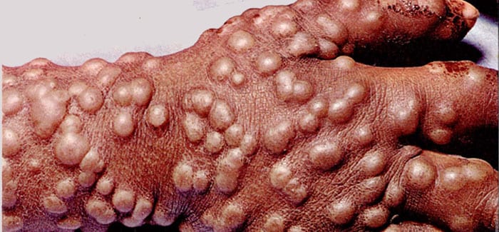
Smallpox vs severe chicken pox:


Smallpox

Virus/Family

Genome

Disease

Transmission/Dx

Tx/Vaccine

Comment

Poxviridae

Smallpox aka Variola

linear dsDNA

URT & 2nd viremia->rash; prodromal fever then centrifugal, regular rash

fomite or lesion/ Guarnieri body

Vaccine available; VIG 1st; Cidofovir 2nd

very contagious, can go through skin; humans only host

chickenpox-

Virus/Family

Genome

Disease

Transmission/Dx

Tx/Vaccine

Comment

alphaherpesvirus

Varicella

envelop linear dsDNA

chickenpox 1/5 childhood rashes

Aerosol

Dx: rash, PCR, culture

high doses of acyclovir; VZIG-> passive immunity

More dangerous in adults due to higher chance of pneumonia

These aren’t even kind of similar, are BOTH VACCINE PREVENTABLE (Hello?) And confusing the two was only an issue before modern medicine. Maybe it’s still confusing to poor Tristan, but to people with basic cognitive functioning, they’re not even close.

Differences-Between-Chickenpox-and-SmallpoxNot the same characteristic rash, not the same clinical presentation, not even the same virus family. In smallpox, lesions begin on the face and then spread outwardly to the extremities, and all lesions will be in the same stage of development. The rash begins at the scalp and head and spreads to involve the trunk and extremities, and the pustules will be at all different stages.

smallpoxchickenpox.jpg


Chickenpox is similar to the same family like HSV, with

Cowdry A or “ground-glass” type inclusions, multinucleated cells, marginated chromatin, and nuclear molding seen under a microscope. Not like smallpox with characteristic Guarnieri bodies.


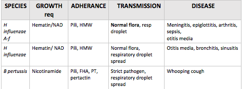
Pertussis vs. H. influenzae

Pertussis and H. influenzae have similar routes of transmission and both cause respiratory infections and otitis media. However, they are also vastly different. Pili is a virulence factor common in gram negative strains that is involved with adhering to host cells, so it’s no surprise that both of them have pili.

Screen Shot 2018-04-20 at 12.23.36 AM


Iron lung vs respirator :

This one is the funniest to me. No shit, we use more respirators today. How is that an argument? It would take him seconds to google “how does an iron lung work?” And another six seconds to google “why the flying fuck would someone with more money pay out their ass for an iron lung if it worked the same as a respirator?” If one cost more, anyone with a functioning brain would obviously pick the much smaller respirator over a giant iron lung they had to stay in a majority of the day. And none of this has anything to do with hiding new diagnoses, considering that makes no fucking sense. Yes, we have better technology now. Respirators and iron lungs do not heal people – they help lung function and prolong lives.

COPD – these are the patients using the respirators you’re thinking of. It’s usually from smoking. No, this isn’t secretly polio either, considering we’re STILL TREATING ELDERLY POLIO PATIENTS (sorry) and chronic bronchitis is an obstructive lung condition that comes with specific criteria that doesn’t involve the diaphragm being paralyzed. Yes, Tristan, I’m positive.


Hepatitis vs the alphabet

– The alphabet is not, in fact, hepatitis. Hepatitis is caused by completely different viruses and is named for their affect on the liver. This isn’t rocket science. HepA is normally acute and is in the same family as Coxsackieviridae. HepB is a herpesvirus. HepC is most likely to turn chronic and is in the same family as Rubella. If you’re confused, use the mnemonic “vowels affect bowels”


Guillain-Barre Ddx:

  • The paralytic form of rabies shares features with Guillain-Barré syndrome but is clinically distinct because of the presence of myoedema, progression to coma, and urinary incontinence
  • Diphtheric polyneuropathy vs Guillain–Barré syndrome (GBS): diptheric polyneuropathy has a slower onset and is more likely to lead to a long term complication or death from diaphragmic paralysis
  • You’re way more likely to get Guillain-Barre from a vaccine-preventable disease than a vaccine


Information sources:

  1. Dabbs, David J. Diagnostic Immunohistochemistry: Theranostic and Genomic Applications. Elsevier, 2019
  2. Dukes, M N. G, and Jeffrey Aronson. Meyler’s Side Effects of Drugs: The International Encyclopedia of Adverse Drug Reactions and Interactions. 16th ed., Elsevier Science, 2015.
  3. Farizo KM, Cochi SL, Zell ER, et al: Epidemiological features of pertussis in the United States, 1980-1989. Clin Infect Dis 1992; 14: pp. 708-719
  4. Goljan, Edward F. Pathology. Saunders/Elsevier, 2014.
  5. Kellerman, Rick D., et al. Conn’s Current Therapy 2018. Elsevier, 2018.
  6. Robbins, Stanley L., et al. Pathologic Basis of Disease. Saunders Elsevier, 2015.


Image sources:

  1. http://bacteriologynotes.com/habitat-and-morphology-of-corynebacterium-diphtheriae/
  2. http://bacteriologynotes.com/laboratory-diagnosis-treatment-and-prevention-of-corynebacterium-diphtheriae/
  3. Diphtheria ECG
  4. Case Study: Faucial Diphtheria Complicated with Myocarditis and …

  5. https://www.visual-science.com/projects/science
  6. Oleksiy Maksymenko, Getty Images
  7. https://hoool.com/an-overview-of-strep-throat/http://jvi.asm.org/content/86/23/12571/F2.expansion.html
  8. http://varicellas.com/2015/03/page/30/
  9. Sketchymedicine.com
  10. https://microbiologyinfo.com/differences-between-chickenpox-and-smallpox/
  11. http://tryptopham.sodoto.net/emrash/
  12. McKinley et al. 1999 Nontraumatic Spinal Cord Injury: Incidence, Epidemiology, and Functional Outcome
  13. https://www.sciencesource.com/archive/Bordetella-pertussis–SEM-SS2576968.html
  14. https://www.slideshare.net/Prezi22/rash-illness-training


Pathophysiology of Vaccine-preventable diseases: Part 2

Viruses are very different. abide to a set of rules:

Rules of Viruses

  • Viruses must replicate to survive.
  • Viruses can’t replicate without a host cell.
  • All viruses replicate using the same basic steps, but the mechanism varies depending on viral makeup. 
    • You and another person may take the same route to work, but one of you is using a bicycle and another is using a car.
  • Viruses need the machinery of the host cell to replicate.
    • And we know that viruses need to replicate to survive.
  • If a protein is vital to the survival of the virus, either the virus genome already encodes it or it will use the host’s code. There are no other options. Examples: polymerase and reverse transcriptase 
    • If it needs something, it’s going to be pre-programmed or you’re going to have it pre-programmed.
  • Larger viruses encode non-vital proteins that help the process of replication. Example: herpes scavenging enzymes 
    • Larger viruses like to encode proteins that help the replication process be more efficient and make less mistakes.

Viruses are weird.  They’re so weird that conspiracies are spread claiming they’re not real, not infective, or man-made. On one hand, I’m not surprised I’m seeing this in the depths of the internet in 2018, considering there are full grown adults who genuinely believe the earth is flat. On the other hand, I can understand their confusion and frustration, as the existence of alive-but-not-alive parasites sounds like exaggerated sci-fi. However, unlike the claims I’m seeing in terms of them being “new,” viruses were here long before modern medicine. They’re not a new, they’re just newly observed and evolve over time to become more infective. Centuries ago, one could still diagnose some viral illnesses based off of the similar clinical presentation and characteristic rashes, but even after Leeuwenhoek’s significant contribution to science, you couldn’t whip out a light microscope to directly observe them. The only viruses that can technically be seen with a light microscope are poxviruses. Even then, you’re going off of tiny inclusion bodies and will not have a detailed view.

While viruses are unique from one another in terms of infectivity, antigens, and appearance, they do share many of the same features. All viruses have nucleic acid and a protein coat called a capsid, which protects that nucleic acid so it can survive in the host and replicate. Naked DNA and RNA are highly susceptible to denaturation and degradation in biological systems. RNA is more finicky than DNA since it has that extra reactive hydroxyl group, which is why we don’t use RNA as the main blueprint for our own bodies and need that RNA “middleman” for transcription and translation. A virus gives less shits than the honey badger, though, in terms of reactivity of nucleic acid and comes in all flavors of single-stranded or double-stranded RNA or DNA. A virus still needs to protect its nucleic acids, so it covers it up in that special capsid coat. This capsid comes in multiple different shapes and sizes, and will either be naked or clothed (called enveloped). Counterintuitively, this envelope makes the virus more susceptible to degradation by heat or acid, making enveloped viruses more susceptible to host defense mechanisms and generally weaker in the enteric (gastrointestinal) tract. In other words,  while some viruses are stronger than others, enveloped viruses will often be destroyed by the stomach if ingested and have a different route they take for infectivity, such as inhalation or sexual intercourse. It doesn’t mean they’re weak viruses, though. One example would be Human Immunodeficiency Virus, or HIV.

The complete virus particle consisting of the nucleic acid and capsid is called the “virion.”

Viral families often share the same general virion structure. For instance, all alpha, beta, and gamma herpesviruses have a pleomorphic icosahedral capsid consisting of 162 capsomers surrounded by an envelope with embedded glycoproteins:

Herpesviridae_virion.jpg
Structure of Herpesviruses (150-200nm in diameter)

This is just a fancy way of saying that they look the same and they’re built with the same general blueprint (so they are related to one another taxonomically), but they act differently from one another in your body and will be recognized differently. In other words, getting infected with genital herpes HSV2 won’t protect you from getting cold sores via HSV1, as they’re not the same virus. So the differences in each virus comes in outer antigens (they’re recognized as different from one another by your body) and pathogenesis.

All viruses have the same general life cycle inside of a eukaryotic host:

Source: Khan Academy

These steps are targeted by antiviral drugs. Of course, the life cycles are a tad more complicated than the general overview shared by all and have a different exit route according to whether or not they have an envelope, which is why one antiviral doesn’t work for every virus (wouldn’t that be nice)


Today we can actually observe viruses infecting cells, incorporating their host genome, packaging proteins, and egress.

Virus packaging materials:


So what viruses do/did we vaccinate against?

1. Smallpox:

Smallpox is the textbook example of vaccine prevention, since we almost completely eradicated it (except for the strains available in labs). What was once a horrible disease now ceases to exist, thanks to the discovery of Edward Jenner.

What did it look like?

Poxviridae_virion.jpg

The capsid had complex symmetry, and the overall structure was brick-shaped.


How was it transmitted? respiratory droplets

Pathogenesis and Clinical Presentation:

It multiplied in the throat and then entered the bloodstream where it multiplied further in lymph. About 2 weeks after infection the characteristic rash would appear all over the body in the same stage of development (unlike chickenpox). The spots developed into vesicles and then pustules. The result was that the patient was either dead or scarred.

How does it appear under a microscope? You’d see viral inclusion bodies called Guarnieri bodies that are typical for all poxviruses

How would you diagnose it, then? Both clinical presentation and diagnostic tests testing specifically for the DNA such as ELISA and PCR with western blotting. You would never ever ever ever ever (continued 100x) have a clinical presentation of smallpox without knowing for certain that the person does or doesn’t have smallpox by running specific diagnostic tests. Unless you’re a fan of losing your job and having the entire country hating your guts.

Characteristic rash:

2. Influenza

Pathogen:

What does it look like?

it’s an enveloped virus with a ssRNA(-) genome. It is pleomorphic with a filamentous capsid

Incubation period: 1-4 days

Spreads via: respiratory droplets

Who is most vulnerable to major complications from influenza?  adults >65 and children<5 years, health risks: immunocompromised patients or patients with CVD, diabetes, and seizure disorders

Pathogenesis:

Influenza A: A bad flu – Pandemic influenza

Influenza B: causes epidemics and is usually less serious than influenza A, though children are badly affected. The deaths from influenza B infection often come from myocarditis as well as bacterial pneumonia

Influenza C: Sporadic flu, less serious URI

Antigenic drift → leads to small variations in the surface proteins.

  • previous exposure means the person will have partial protection, causing a milder disease.

Antigenic shift → viruses that infect different species infect the same cell and create new viruses during replication with mixed HA and NA antigens.

  • Of the three types of influenza virus (types A, B, C), only influenza type A infects multiple species and hence is the only type in which this occurs.
  • The different HA and NA antigens are given a number subscript, for example H5N1 (avian influenza, or “bird flu”). These new strains, to which no one is immune at first, are what
    cause pandemic flu.

Complications:

  • Most who are immunocompetent will be able to fight off influenza.
  • Bacterial coinfections of S. aureus (“staph”), S. pneumoniae, S. pyogenes, or H. influenzae occur in approximately 40% – 90% of fatal influenza virus infections.
  • encephalopathy,
  • transverse myelitis
  • Reye syndrome
  • myositis,
  • myocarditis
  • myocardial injury

Signs/Symptoms:

children— Otitis media, nausea, vomiting, and myalgias  (usually soleus and gastrocnemius)

Adults and children: abrupt onset of fever, myalgia, headache, malaise, nonproductive cough, sore throat and rhinitis.

How does the influenza vaccine work?

There are two ways in which ρ epitope can be used to improve vaccine development. The first is the development of “like” strains, where the value of p epitope  where a similar strain is chosen from several possibilities. It can be used to quantify how close each of the “like” strains is to the desired vaccine strain. This method is difficult, as it is practically impossible to produce in large quantity the exact strain of influenza that is desired for the vaccine, especially in multiple locations across the globe.

The second way in which p epitope can be used is identification of the strains desired to be included in the vaccine. That is, given a list of potential circulating strains, each with probabilities of outbreak for the upcoming year, which vaccine strain minimizes the weighted distance from the potential circulating strains? The value of pepitope can be used to define distance, and so to help choose the closest vaccine strain to the potential circulating strains.


3. Varicella zoster

What does it look like? A typical herpes virus –  an enveloped double-stranded DNA virus with icosahedral capsid

How does it spread? Through respiratory droplets and cutaneous lesions. Varicella is a highly contagious, affecting 90% of a family within two weeks if one member gets sick. Community outbreaks usually occur in late winter and early spring months in temperate regions. Unlike chickenpox, Herpes zoster (“shingles”) is nonseasonal. Before vaccination, it was also a common cause of nosocomial outbreaks 

Incubation period: averages about 2 weeks – can range from 11-20 days after initial exposure

Pathogenesis:

The virus disseminates into skin and mucous membranes, developing the characteristic lesions. Dissemination may spread to other sites, such as the lungs, in immunocompromised individuals. Adults tend to have more severe clinical presentations compared to children. After infection it remains latent in the posterior dorsal root ganglia.


Clinical presentation:

The rash begins at the scalp and head and spreads to involve the trunk and extremities. Lesions progress through specific stages of erythematous macules, vesicles, pustules, then crusts.


Complications:

Complications in children are common in the immunocompromised. This includes continued lesion development, a very high fever; and visceral involvement. The visceral involvement includes pneumonia (most common complication), meningoencephalitis, and hepatitis. 


Untitled picture

Adults with a normal immune system typically present with viral pneumonia – a complication that is 25-fold higher than in children. This develops about 1 – 6 days after the onset of rash. Symptoms include:

  • cough, shortness of breath, chest pain, and coughing up blood. 

Pregnant women also experience pneumonia, and varicella is known to cause spontaneous abortion from fetal demise or birth complications such as growth restriction and musculoskeletal abnormalities

Fetal death from varicella zoster at 26 weeks
Fetal liver destroyed by varicella zoster

.

Congenital varicella syndrome

4. Rubella

What does it look like?

It’s an  enveloped virus of the togaviridae family with +ssRNA

How does it spread? Through respiratory droplets

Pathogenesis and clinical presentation: Patients present with a rash and arthritis

Screen Shot 2018-04-19 at 11.42.35 PM

It is a known ToRCHeS infection – it is teratogenic, which means it crosses the placenta and causes fetal damage.

Screen Shot 2018-04-19 at 11.54.42 PM
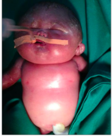
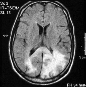
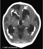
If the fetus survives, a neonate with congenital rubella will present with the characteristic cataracts, blindness, deafness (most common), thrombocytopenia, heart defects, hepatosplenomegaly, and skeletal deformities. There will also be a large amount of IgM in cord blood. The childhood presentation often consists of a fever with lymphadenopathy and rash and sometimes a secondary spread to CNS consisting of progressive rubella panencephalitis. In adults it may present with post-infectious encephalomyelitis and optic neuritis, myelitis, Guillain Barre syndrome.

5. Measles:

What does it look like:

It’s a ssRNA(-) virus of the paramyxoviridae family


How does it affect people?

The measles is the hallmark “it’s just a childhood illness” of anti-vaxxers. They’ll continue to defend their “childhood illness” because it makes them feel less shitty for being personally responsible for every outbreak since the vaccine was introduced.

Them: “Oh yeah, well you vaxxers shed!”

Except vaccinated kids don’t shed the measles. If anti-vaxxers ever stopped to read the vaccine inserts (they never do) rather than talking about it, they would know that the MMR vaccine insert is about 20 pages, not a screenshot downloaded from some crunchy granola site, and specifically states that it doesn’t shed. Whoops!

Them: “Well MMR doesn’t protect very long”

It actually protects for a long time, and anyone can get a booster if they’re concerned. Anti-vaxxers seem to be convinced that no one gets boosters. We do. And MMR is efficient.

The measles vaccine alone saved 15.6 million lives annually worldwide annually from 2000-2013. Think one vaccine can’t make a difference ? Think otherwise. Just one vaccine saved many lives in Maputo: https://www.ncbi.nlm.nih.gov/pmc/articles/PMC2486532/

Most importantly, it’s not “just a childhood illness,” it’s extremely contagious and deadly. Japan discovered this the hard way. After taking MMR out of the required childhood vaccines, Japan had 200,000 cases of the measles and 88 deaths in just the year 2000 alone: http://www.thelancet.com/journals/lancet/article/PIIS0140673604167159/fulltext

This fit in perfectly with expectations from older peer reviewed science where they first started noticing a trend: https://www.med.nagoya-u.ac.jp/medlib/nagoya_j_med_sci/5514/v55n14p23_32.pdf). Herd immunity is determined via math, so it’s not surprising that they could predict a trend. Japan has been so affected by the measles that they lost a reported $404 million to measles treatment in comparison to vaccination, which costs $165 million https://www.ncbi.nlm.nih.gov/pmc/articles/PMC3217873/ Normally it costs the government money to vaccinate (I get mine for free), but not paying for so many measles-induced hospitalizations would had saved them hundreds of millions of dollars. And it was all for nothing. Regardless of fear-mongering, the autism rate in Japan continued to rise as MMR went down: http://onlinelibrary.wiley.com/doi/10.1111/j.1469-7610.2005.01425.x/abstract

Another common antivaxxer myth:

Measles can be cured with Vitamin A!” (e.g “No doctor wants to tell you…”)

Vitamin A can potentially help shorten the duration of the illness and even stop measles progression in many circumstances, and this isn’t some special secret. I’ve never met a single doctor who was “anti-vitamin A.”

Here are the problems:

  1. Why expose yourself to the measles toxin when we have a safe way to prevent it?
  2. Taking copious amounts of vitamin A is toxic, as it’s a fat soluble vitamin. This can lead to cerebral edema and liver damage.
  3. It still doesn’t mean you’re safe. Vitamin A isn’t a cure, but it is recommended for those who have just been diagnosed.

Personally, I prefer prevention rather than simply tossing drugs at people, as anti-vaxxers seem to prefer. Overall, vitamin A can aid in the progression of measles as any healthcare professional will tell you, but it is not a “proven cure,” and is a fat-soluble vitamin, meaning you don’t simply excrete it in the urine like you would with vitamin C. It can be toxic. Someone giving their child a ton of vitamin A in attempts to cure their measles without the help of a medical professional could subsequently kill them. It makes a lot more sense to simply not get the measles and also not contribute to increasing cases of the measles.

But if Vitamin A helps the measles, that means the measles is a nutritional deficiency!

This seems to be a popular idea among those who are anti-Germ Theory (I still laugh typing that out). That’s not how the immune system works. Eating healthy and exercising can help your immune system function well, but it’s not a cure. Any doctor out there will ask their patients to eat healthy and exercise. This isn’t a secret either. Back in the “good old days,” we didn’t have processed food. Think about the logic there if it’s supposedly a nutritional deficiency.

Anti-vaxxers: “Well, it’s so rare in the United States it’s not like my child is at risk anyways!”

Your unvaccinated special snowflake is actually 35x more likely to contract the measles compared to their vaccinated friends. When measles was prevalent in the pre-vaccine era, most people experienced infection by the age of 20, and 90% of these reported cases were seen in those kids younger than 10 years old. Your child is highly at risk of contracting the measles.

So how does it spread so easily? It spreads through small-particle aerosols and, unlike many other viruses, can remain infectious for several hours at low relative humidity. It has caused secondary infections in the absence of face-to-face contact with an index case.

Incubation period: about 9-14 days (may be longer in adults)

Fatality: Lower in developed countries and as high as 25% in some developing countries. Most of these deaths result from respiratory tract involvement and/or neurologic complications.

Pathogenesis and Clinical presentation:

Spreads through the respiratory tract and consists of two stages: Viremic and a second phase of viremia that’s either prodromal or involves dissemination. The disseminated phase can involve the respiratory tract, gut, bile duct, and bladder and spread to lymphoid organs. The typical measles virus–induced giant cells may be present in the tonsils, appendix, other lymphoid organs, and various epithelial surfaces, including those of the respiratory tract. In a typical case, symptoms start similar to a cold. Photophobia (sensitivity to light) usually develops by the second day. Soon, Koplik’s spots develop, small ~2mm macules with a central white dot, will be found around the area of the parotid duct. By the fourth day, these are usually gone and an erythematous rash develops behind the ears. This becomes maculopapular and spreads from the face down the body over the trunk and libs. The fever will increase. The person will typically get better a few days later, but a slew of complications can develop such as an ear infection, pneumonia, encephalitis, subacute sclerosing pan-encephalitis (SSPE- a horrible progressive inflammation in the brain), diabetes, and thrombocytopenic purpura. The most common cause of death in young children is from the measles is pneumonia, which affects about 1/20 of those who contract it. 1/1000 develop SSPE, especially in cases involving younger children. For every 1,000 children who get the measles, 1-2 will die. What can help the measles? Vaccination. Before vaccination, there were about 50 cases of SSPE annually. Now there are only 1 or 2 cases annually.

Pasted Graphic 3 Pasted Graphic 6

Important facts about the measles: 

  • As many as one out of every 20 children with measles gets pneumonia, the most common cause of death from measles in young children.
  • About one child out of every 1,000 who get measles will develop encephalitis (swelling of the brain) that can lead to convulsions and can leave the child deaf or with intellectual disability.
  • For every 1,000 children who get measles, one or two will die from it.

Information sources:

  1. Dabbs, David J. Diagnostic Immunohistochemistry: Theranostic and Genomic Applications. Elsevier, 2019
  2. Dukes, M N. G, and Jeffrey Aronson. Meyler’s Side Effects of Drugs: The International Encyclopedia of Adverse Drug Reactions and Interactions. 16th ed., Elsevier Science, 2015.
  3. Farizo KM, Cochi SL, Zell ER, et al: Epidemiological features of pertussis in the United States, 1980-1989. Clin Infect Dis 1992; 14: pp. 708-719
  4. Goljan, Edward F. Pathology. Saunders/Elsevier, 2014.
  5. Kellerman, Rick D., et al. Conn’s Current Therapy 2018. Elsevier, 2018.
  6. Robbins, Stanley L., et al. Pathologic Basis of Disease. Saunders Elsevier, 2015.


Image sources:

  1. http://bacteriologynotes.com/habitat-and-morphology-of-corynebacterium-diphtheriae/
  2. https://sonoworld.com/Fetus/Case.aspx?CaseId=604&answer=1
  3. http://bacteriologynotes.com/laboratory-diagnosis-treatment-and-prevention-of-corynebacterium-diphtheriae/
  4. Diphtheria ECG
  5. Case Study: Faucial Diphtheria Complicated with Myocarditis and …

  6. https://www.visual-science.com/projects/science
  7. Oleksiy Maksymenko, Getty Images
  8. https://hoool.com/an-overview-of-strep-throat/http://jvi.asm.org/content/86/23/12571/F2.expansion.html
  9. http://varicellas.com/2015/03/page/30/
  10. Sketchymedicine.com
  11. https://microbiologyinfo.com/differences-between-chickenpox-and-smallpox/
  12. http://tryptopham.sodoto.net/emrash/
  13. McKinley et al. 1999 Nontraumatic Spinal Cord Injury: Incidence, Epidemiology, and Functional Outcome
  14. https://www.sciencesource.com/archive/Bordetella-pertussis–SEM-SS2576968.html
  15. https://sonoworld.com/fetus/page.aspx?id=153
  16. http://www.cmaj.ca/content/168/5/561?utm_source=TrendMD&utm_medium=cpc&utm_campaign=CMAJ_TrendMD_1


Pathophysiology of vaccine-preventable diseases: Part 1

Every now and then, I’ll scroll through my feed to see a giant red headline that has the word “truth” flashing at me somewhere in the title, such as  “Truth Doctors Won’t Tell You About ‘Deadly’ Childhood Diseases!” The article’s overall point will be scoffing at the notion of acquired infections, claiming how they’re simply childhood illnesses overdramatized by Big Pharma, without noticing the coincidental nature of presenting this information through histrionics and false statements. One of the funniest overused tactics to me is a baby covered in needles, as if doctors are treating children like tiny human pincushions:

I’d be terrified, too, if a bunch of needles were simultaneously injected into various places in my arm with no warning or reason. Most parents have seen their baby get vaccinated at this point, and those of us on planet earth know it looks like this, instead:

Okay, the baby being calm and collected while they get vaccinated might be a tad exaggerated.

Sticking multiple needles into a child as a fear-tactic is like taking anti-vaxxers’ Starbucks cups over 18 years, pouring the gallons in a giant jug, and claiming that’s how much coffee they get on the regular. Not to mention, most of us without aichmophobia are well aware that diseases are scarier than needles. I know a number of people personally who frequently use needles for their chronic conditions. Needles are sometimes used even for family planning.

Here’s a realistic look at the difficulties many experience while trying to conceive through vitro fertilization:

source                                                                               source

Want to include all evil drugs by Big Pharma and not just injections?

Anti-vaxxers, meet the reality of cystic fibrosis:

Source

I don’t think I’m met a single person with a terminal condition who is anti-vaccine. While there’s a real world of chronic illness, pain, and deadly infectious diseases, there are still affluent families that remain clueless and whip out headlines like this:

Screen Shot 2018-04-04 at 11.48.49 PM

Congratulations. Gold star for you. Other moms are.

I also have a feeling that if I click on most of these headlines, my computer will end up with a greater number of viruses than one would acquire sharing needles at Woodstock, which is perhaps the point they were trying to make. “See you could get your computer fixed! I’m sure your body will be fine!” It is both horrifying and amazing that we live in a time that has progressed so much in the field of medicine that rich white parents in suburban California are now willingly choosing to avoid vaccination because they’ve been privileged enough to never witness it.

I’ve only personally experienced the “typical childhood” disease, chickenpox, while just a century ago parents were burying their eighth child from diphtheria after watching them slowly suffocate and die without having any control over the situation. The difference is being born later. I consider myself extremely lucky, though I’m not looking forward to the shingles.


You may have heard that kids are exposed to more pathogens in one day then they’ll receive in a lifetime of vaccination. This is true. Kids who aren’t born with a primary immune deficiency such as X-linked SCID can certainly handle antigens. Our immune systems can recognize 10 7 –10 9 different antigens, which is mind-boggling to me. That’s a lot more than you’ll ever get in any vaccine.

Antigens, in terms of vaccination, are recognizable and  unique parts of a specific bacteria or virus that your immune system uses as a blueprint to create new antibodies. The next time your body is introduced to the organism, through either a subsequent/booster vaccine or the real deal, you will fight it quicker and more efficiently. After vaccination, the antigen is picked up by antigen presenting cells (APCs). APCs will ingest the antigen and vomit tiny bits of it out to display it on tiny microscopic kabob. Dendritic cells, a type of APC, will migrate to the draining lymph nodes to present the antigen to T cells. Antigens can also be brought to draining lymph nodes by subscapular macrophages to the B-cell zone. Either way, even if antigen is presented to T cells, most vaccines (especially killed vaccines) have a tendency to influence humoral more than cell-mediated immunity.

Bacteria are prokaryotic organisms, which differ from our cells (eukaryotic). Bacteria are tiny unicellular organisms that reproduce a lot quicker than we do, have different wall structures, and don’t have membrane-bound organelles. Most of the time they use our body as a host to grow and flourish. Other times they try to invade. We even use their features to our advantage at times. I use E. coli all  the time in the lab. E. coli is chosen as the staple bacterium in a microbiology lab because they’re the rabbits of the bacteria world. They can double the population in 20 minutes. They breed so quickly that they can technically populate faster than they can replicate.

Bacteria not only have their host genome, but they have a second set of small, circular DNA called a plasmid. This plasmid is like a set of game cheat codes. Through the plasmid they get virulence factors, resistance, and sex pili.

Bacteria have been evolving for a very, very, very long time. I was taught incorrectly about evolution growing up, and I always associated evolutionary advancement with large, complex beings like humans. Bacteria are also evolutionarily advanced. They’ve been here longer than we have. Over time pathogens that infect humans have developed ways to evade our body’s defenses. Sometimes they have virulence factors that directly harm the host. Other times they’ll hide inside of our own cells. Sometimes they’ll even release signals to our cells that calm down our immune system. We’ve seen their ability to evade antibiotics since the discovery of penicillin.


So why do we vaccinate? Because vaccination presents the antigen without the infection. It’s like giving your immune system a 3D print to use for antibodies. You have many different types of antibodies, but the one that is released upon first exposure to an organism is IgM, which is a very large and is made up of five different antibodies bound together. IgG is released upon second exposure, and is one antibody. It is the type of antibody that is tiny enough to cross the placenta as well. IgG means a faster, more efficient antibody response with more evolvement of the adaptive immune system. So when you’re exposed to the antigen for real, your body already knows how to fight it.


What bacteria have available vaccines in the US?

1. H. influenzae

Why it’s so dangerous:  Young children under the age of 2 are particularly susceptible to H. influenzae, and this form of meningitis can cause death in less than one day.

Possible confusion: some may be confused between serotypes of H. influenzae. Serotype B is the encapsulated serotype known for causing meningitis. Other serotypes can still be dangerous by causing bronchitis/pneumonia, but are more likely to cause otitis media.

Why are encapsulated organisms more likely to cause meningitis? 

Activators of the alternative compliment system are not in CNS. The compliment system is a cascade of natural host responses to combat an invading pathogen. It can use a classical pathway, but sometimes involves other alternative pathways, but with the same overall effect. The compliment system involves signaling other immune cells to come help out, neutralizing the pathogen so it’s less toxic, labeling the pathogen to be destroyed, and the final step of literally poking a hole in the organism called the “membrane attack complex.” Organisms that have a capsule are susceptible to the membrane attack complex. Think of a medication capsule:

This capsule prevents the organism from being swallowed by macrophages (a type of APC). Once you poke a hole in most encapsulated organisms, they’re not going to stay around to breed — they get eaten. They’re pretty weak without that capsule. However, one’s CNS doesn’t have the same complement system, so the encapsulated bacteria that survive the rest of the body will then be able to wreak havoc on someone’s central nervous system. It’s bad enough for adults, much less in toddlers.


Pathogenesis:

The infection can be endogenous or exogenous. This means that it can start from either inside your body or you contract it from another sick individual. So H. influenzae is often found as part of the normal flora of the upper respiratory tract. It’s not a rare bacteria at all.  And just like your roommate, sometimes organisms that live with you decide one day to stab you in the back. Bacteria in the pharynx invade the lymph/blood and then travel to the meninges. The bacteria will spread throughout the central nervous system and sometimes take a detour to bones and joints.

The power of vaccination: 

  • Vaccination has reduced invasive disease rates by 99%
  • Before vaccination, 1/200 children used to develop pertussis by the age of 5. Those who are most susceptible are young children <2 y.o, as this is the age most likely to develop meningitis. Children 2-5 are more likely to develop symptoms of epiglottitis and pneumonia
  • The risk of serious infection for unvaccinated children younger than 4 years of age living with an index case is more than 500x that of unexposed children. This risk indicates a need for protection of susceptible contacts
“The decline in Haemophilus influenzae type b (Hib) meningitis in association with the introduction of new vaccines is shown. Note also the steady state of the other major causes of childhood meningitis; they did not increase to “fill in the gap” nor did H influenzae invasive disease caused by other serotypes.”

Evolution of the H. influenza vaccine: If vaccines were all about money in a giant government conspiracy, we wouldn’t be seeing the same government losing money in grants for a better alternatives for vaccines that already exist. H. influenza is no exception to this. The first vaccine, a purified PRP vaccine, came out in 1985. Why did they change it? The immune system of infants didn’t create the desired cell-mediated immune response, so it was only slightly effective, and the age group that had the lowest immune response was also the age group particularly susceptible to contracting meningitis. So a new protein conjugate vaccine was created for use in 1989 that linked PRP to proteins that would create an immune response. This became the universal vaccine  by the end of 1990.

Life before vaccination: 1/200 children used to develop Hib by the age of 5
MC invasive form: meningitis, usually <2 y.o
Children 2-5: MC epiglottis and pneumonia
meningitis


2. Diphtheria

Pathogen: Bacteria- Corynebacterium diphtheriae

What does it look like?

  • consists of a non-motile, non-capsulated, non-
    spore-forming aerobic bacillus.
  • Gram-positive, but easily decolourized during the staining procedure
  • On microscopy, C. diphtheriae exhibits considerable pleomorphism, setting it apart from other species
  • The arrangement of organisms on a smear often resembles “Chinese letters,” which are unique to diphtheriae

INCUBATION PERIOD: 2–5 days

WHO IS MOST AT RISK? Diphtheria tends to affect young children

How is it diagnosed?

Diphtheria is detected by culture and simple screening tests, as pathogenic diphtheriae produce cystinase instead of  pyrazinamide. This will be seen as a brown halo around colonies in a modified Tinsdale’s agar. This is all just a very fancy way of saying that diphtheria produces a type of enzyme that allows us to diagnose it if we use agar with special chemicals that make the action of the specific enzyme stand out with a brown color.

Signs often include: pseudomembranes, foul odor, lymphadenitis, edema (depends on type)

OTHER UNIQUE SIGNS/SYMPTOMS – a result of the exotoxin’s effects on epithelial cells.

HALLMARK OF DIPHTHERIA: The grey-white pseudomembranes (similarly to what is seen in the colon with a C. diff infection)

The pseudomembrane is made up of cell debris and fibrin along with leukocytes and bacteria. It adheres to underlying tissues, but bleeds if you try to remove it. The disease is classified according to location.


How does diphtheria kill, and why are we so concerned about it, if it’s just a swollen neck and really bad breath?

Because of the Diphtheria exotoxin. This is a a 62 000 Da polypeptide (a fancy way of saying a protein) that includes two segments “A” and “B”:

  • A- this is the active toxin
    • This catalyzes a reaction that inactivates our tRNA in that cell by binding to something called “elongation factor 2” (EF-2)
      • EF-2 is needed to transfer codes from mRNA to tRNA to make proteins
      • If it’s inactivated, it stop protein sythesis
  • B- this is the binding segment that allows toxin A to enter the cells by binding on the membrane

complications of the exotoxin:

  • The most common toxic complications of diphtheria are myocarditis  and neuritis.
  • Malignant diphtheria has a mortality rate as high as 50% during the acute phase
    • they are likely to die from the remote effects of the toxin. For example, sudden cardiac death can occur up to 8 weeks following the acute disease.
    • The bullneck is not just a typical presentation of diphtheria, but it’s also a huge predictor of the development of myocarditis, which takes place in 2/3 of severe cases.
      • The mortality rate of diphtheritic myocarditis is about 50%.
  • 10–20% of cases lead to neurotoxicity.
    • These complications develop ~3-8 weeks after symptoms first start. It often begins with paralysis of the soft palate (the roof of your mouth that is softer and closer to your throat), which will present itself with a “nasally” voice and regurgitation of fluids through the nose.
    • Paralysis continues, leading to blurred vision and cranial nerves IXth, Xth, VIIth greatly affected. Unlike other forms of paralysis, Neurological deficits seem to be disorganized, with cranial nerve deficits improving while peripheral nerve deficits worsen.When neurotoxicity develops, this often leads to quadriparesis and death from respiratory failure from paralysis of respiratory muscles the throat (larynx) closing.
      • Limb weakness occurs in about 50% of those with neuropathy.
      • Sensory deficits and autonomic dysfunction are other common manifestations
    • 7–10% of moderate/severe cases develop polyneuritis.
    • The exotoxin will lead to segmental degeneration of the myelin sheath. In severe cases, axons will degenerate.
    • Unlike Guillain–Barré , as high as 41% of those with diphtheric polyneuropathy still couldn’t work a year after contracting the disease. Antitoxin is also largely inaffective in preventing neuropathy


3. Whooping cough

Pathogen: Bordatella pertussis

What does it look like? It’s a small, aerobic (it survives in lots of oxygen which is why it likes the respiratory tract), gram-negative rod. 

How is it spread? Through respiratory droplets. It’s extremely contagious, and about  80% of people in a household will contract it if one person is affected

Who is most at risk? The population most at risk from dying of a pertussis infection are unvaccinated infants younger than 6 months of age

Why is vaccination important? According to epidemiological studies, It occurs most commonly in nonimmunized or only partially immunized children, and a large percentage of individuals have to be vaccinated for herd immunity to take place. It’s is one of the leading causes of vaccine-preventable deaths worldwide. Immunity to the “natural” B. pertussis infection anti-vaxxers boast about isn’t permanent. Vaccination extends immunity, protects the community, and prevents the illness.

Speaking of epidemiological studies, how often do people die of whooping cough annually? There ~16 million pertussis cases and ~195,000 deaths in children annually.  WHO has estimated that global vaccination against pertussis in 2008 prevented about 687,000 deaths. When vaccination rates drop, pertussis increases. There were about 48,277 reported cases in 2012 in the United States, which sparked education about vaccines. Thankfully, reported cases dropped to about 20,762 in 2015.

Pathogenesis and Clinical presentation

Whooping cough involves a lot of toxins and virulence factors. These include:

pertussis toxin (PT): Disrupts signal transduction, which instigates lymphocytosis, affects  insulin secretion, and enhances the host’s sensitivity to histamine

tracheal cytotoxin: Consists of a sugar and protein derived from their cell well

  • kills respiratory epithelium with IL-1 and nitric oxide (an inflammatory signaling molecule and a vasodilator)

Filamentous hemagglutinin (FHA) and agglutinogens: Help the bacteria attach to the throat

adenylate cyclase: ↑ cyclic adenosine monophosphate (cAMP)

  • This results in inhibition of APC function (specifically neutrophils) and apoptosis (programmed cell death)

pertactin (PRN):

  • An adhesin with a similar action to adenylate cyclase, inhibiting APC function and allowing cell attachment


The bacteria first attach to the epithelium of the respiratory tract, produce toxins that will paralyze the motile cilia, which interferes with clearing mucous and other respiratory secretions, leading to a persistent cough  and inflammation. B. pertussis can evade host machinery by promoting lymphocytosis while impairing chemotaxis. This means you’ll produce many immune cells, but they’ll have trouble getting to the site they need to, which increases inflammation as well as “distracts” the body from fighting the infection. The bacteria can also hide in macrophages that hang out in your alveoli.

There are also specific steps to a pertussis infection: inoculation → attachment → Dividing cells and producing virulence factors → evading host defenses → tissue damage → clearance of disease, chronic illness, or death

It’s called the “hundred day cough” for a reason. There is also a specific clinical course of whooping cough that consist of the following:


  • Incubation period: usually 7 to 10 days, but can range from 4 to 21 days.
    • The person is asymptomatic
  • Catarrhal: (1-2 weeks)
    • The onset is typically insidious and is usually assumed to be a cold
      • Sneezing, fever, rhinitis, mild cough
  • Paroxysmal stage: 1 to 6 weeks, but can persist up to 10
    • A person is most contagious during this period
    • Presents as frequent coughing followed by characteristic “whoop” sound
    • Vomiting and passing out
  • Convalescence: can range from weeks to months
    • Subsequent respiratory infections are common


Diagnosis: Nasopharyngeal culture is the gold standard

Treatment: Vaccination is the most important factor for prevention. Treatment may shorten the duration of the course if treated early – usually macrolides. Otherwise, if it’s caught too late, there’s not much that can be done other than treating symptoms and waiting

Complications: Whooping cough often leads nutrient loss from loss of appetite, otitis media, and dehydration. The most common serious complication of a pertussis infection is pneumonia. Pneumonia can either be caused by B. pertussis infection itself or co-infection with other respiratory pathogens – usually RSV.  In infants, the hypoxia caused by frequent bouts of oxygen deprivation from coughing along with the toxins can lead to neurologic complications such as seizures and encephalopathy. Complications can also stem from high pressure.. This includes collapsed lungs, serious nosebleeds, brain bleeds, hernias, and rectal prolapse.

Here’s a real look at whooping cough: 


 Information sources:

  1. Dabbs, David J. Diagnostic Immunohistochemistry: Theranostic and Genomic Applications. Elsevier, 2019
  2. Dukes, M N. G, and Jeffrey Aronson. Meyler’s Side Effects of Drugs: The International Encyclopedia of Adverse Drug Reactions and Interactions. 16th ed., Elsevier Science, 2015.
  3. Farizo KM, Cochi SL, Zell ER, et al: Epidemiological features of pertussis in the United States, 1980-1989. Clin Infect Dis 1992; 14: pp. 708-719
  4. Goljan, Edward F. Pathology. Saunders/Elsevier, 2014.
  5. Kellerman, Rick D., et al. Conn’s Current Therapy 2018. Elsevier, 2018.
  6. Robbins, Stanley L., et al. Pathologic Basis of Disease. Saunders Elsevier, 2015.


Image sources:

  1. http://bacteriologynotes.com/habitat-and-morphology-of-corynebacterium-diphtheriae/
  2. http://bacteriologynotes.com/laboratory-diagnosis-treatment-and-prevention-of-corynebacterium-diphtheriae/
  3. Diphtheria ECG
  4. Case Study: Faucial Diphtheria Complicated with Myocarditis and …

  5. https://www.visual-science.com/projects/science
  6. Oleksiy Maksymenko, Getty Images
  7. https://hoool.com/an-overview-of-strep-throat/http://jvi.asm.org/content/86/23/12571/F2.expansion.html
  8. http://varicellas.com/2015/03/page/30/
  9. Sketchymedicine.com
  10. https://microbiologyinfo.com/differences-between-chickenpox-and-smallpox/
  11. http://tryptopham.sodoto.net/emrash/
  12. McKinley et al. 1999 Nontraumatic Spinal Cord Injury: Incidence, Epidemiology, and Functional Outcome
  13. https://www.sciencesource.com/archive/Bordetella-pertussis–SEM-SS2576968.html